Meiji Techno, MT5000DSS1, Digital Slide Scanner Imaging System, 1 Slide Scanning
MT5000-DSS1 is Meiji Techno’s complete turnkey digital brightfield imaging platform which allows you to convert your single glass slides into digital data.
MT5000-DSS1 is a complete imaging platform which is perfect for most biological research, educational and industrial applications. It creates high quality digital slides to view a panoramic image of complete slide area. Equipped with a state of the art imaging system, you will get high resolution images due to high quality Japanese optics and optimum LED illumination to produce superior signal-to-noise ratios. It has user-friendly image processing and analysis software for ready to view, diagnose or publish high quality images.
The MT5000-DSS1 has automated single slide analysis with high performance and digital imaging right at your lab bench. Its time saving automation such as auto focus and rapid stage control and automated routines will help reduce time to complete tedious slide scanning allowing high throughput, high data quality transmission and improved laboratory experimental reproducibility.
This system has been designed with advanced capabilities to simplify demanding slide based imaging applications so you can focus on acquiring images and data easily saving you time from instrument manual operation. All systems come with high sensitivity color camera optimized for quantitation and dedicated to high resolution images. There is also an optional software package offering image acquisition and classification tools for your laboratory applications.
Dedicated installation and training program will get your laboratory easily set-up. Our dedicated MT5000-DSS experts will come to your site to provide hands on workflow training and make sure your laboratory is quickly trained to utilize the complete Digital Imaging Platform to maximize your productivity.
Meiji Techno’s MT5000-DSS1 allows you to convert your glass slides into digital data with a complete turnkey system
Features:
- Scanning slide and creating virtual sample
- Automated Focus: automatic, range, dynamic, extended
- Plan Apochromat Infinity Corrected Objectives Immersion scanning with high definition (optional)
- Motorized encoded X and Y Scanning Stage
- Z-Stack Capability
- Built-in Koehler illumination, LED
- Vision Slide®Assist Software
- Automated and manual calculation of optical and geometric parameters of a selected object. Tools to create marks and comments on the digital sample.
- Database for Archive Management
- AutoDetection of scanning area
- Adjustable size of scanning area
- Working with images: Select, Edit, Marks and Text Comments
- Comments to digital sample image
- Education and professional development
- Telemedicine and remote consultations
- Remote access (optional)
- Quintuple Reverse Nosepiece Turret
- Manual slide handling/loading and identification
- Automated single slide scanning 1.5 mins/ Slide (15x15mm)
- Simultaneous loading for single slide applications
- Microscope slide requirements Width 25.26mm, length: 75-76mm, thickness: 0.8-1.4mm
- Automated Focus: automatic X, Y, Z Scanning, extended, range
- Plan Apochromat Infinity Corrected Objectives 20x/0.75- 0.5/0.25 um/pixel.
- Plan Apochromat 40x/0.95-0.25/0.12um/pixel ( Optional)
- Immersion scanning with high definition (optional) LED light source that provide more environmentally friendly solution and more energy efficient
- 110V-240V/ 50-60 Hz
Biological, life science, pharmaceutical and education
- Visualization and storage of digital samples in the database
- A professional set of tools to work with digital samples: create, edit, organize, classify and comment
- Calculation of geometric parameters in standard measurement units
- Automated and manual calculation of optical and geometric parameters of a selected object. Tools to create marks and comments on the digital sample
- Remote access and network capabilities
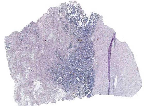
Visualize Panoramic images of the complete slide area
Creation of high quality digital slides. Exceptional quality, high definition and accurate color rendition thanks to Plan Apochromat objectives.
Immersion scanning with high magnification and high definition
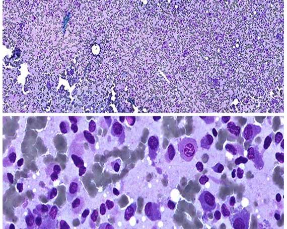
Sample scanning with high magnification (60x, 100x), when the high quality of images is required. For example, for blood and bone marrow samples.
Optional Special high definition objectives are used for immersion scanning. These objectives work with immersion oil.